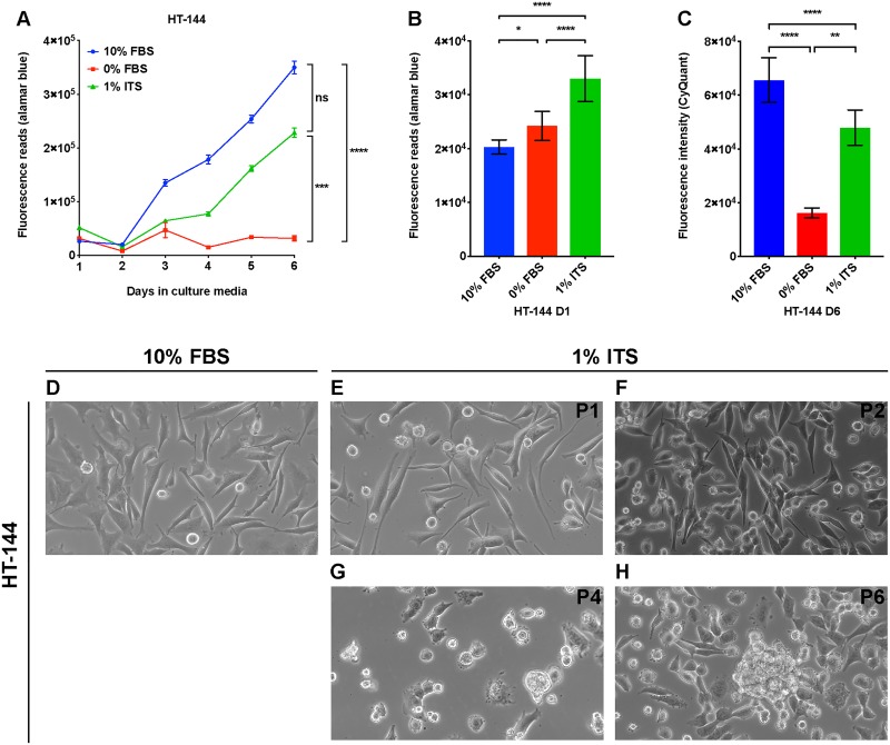
Fig 1. A lipid-free and insulin-supplemented medium supports proliferation of HT-144 melanoma cells.
(A) HT-144 cells were seeded in 10% FBS medium at day zero. On day one, cells were washed with PBS and changed to the indicated medium conditions. Cell proliferation was measured with alamarBlue assay daily for six days. Each data point represents the mean ±SD of quadruplicate samples. The results were analyzed using two-way repeated measures ANOVA followed by post hoc Tukey’s multiple comparison tests. For culture time, F = 1505, P < 0.0001; for culture condition, F = 2153, P < 0.0001; for interaction between culture time and condition, F = 409.4, P < 0.0001. (B) HT-144 cells were seeded in 10% FBS medium at day zero. On day one, cells were washed with PBS and changed to the indicated medium conditions. alamarBlue assay was performed on the cells cultured in the indicated medium for one hour. Results were analyzed using one-way ANOVA followed by post hoc Tukey’s multiple comparison tests. F = 202.7. Significant differences between medium conditions are indicated as *P < 0.05, **P < 0.01, ***P < 0.001 and ****P < 0.0001. ns, not significant. (C) HT-144 cells were seeded in 10% FBS medium at day zero. On day one, cells were washed with PBS and changed to the indicated medium conditions. CyQuant assays were performed on the cells cultured in the indicated medium at day six. Each data bar represents average measurement of five replicate samples. Results were analyzed using one-way ANOVA followed by post hoc Tukey’s multiple comparison tests. F = 80.77, P < 0.0001. Significant differences between medium conditions are indicated as *P < 0.05, **P < 0.01, ***P < 0.001 and ****P < 0.0001. ns, not significant. (D—H) HT-144 cells were cultured in RPMI medium with 10% FBS or 1% ITS supplement for multiple passages. Morphologies of cells cultured in 1% ITS medium from passage one (P1) to passage six (P6) were monitored by light microscopy with 40 × objective and 10 × ocular lens.

