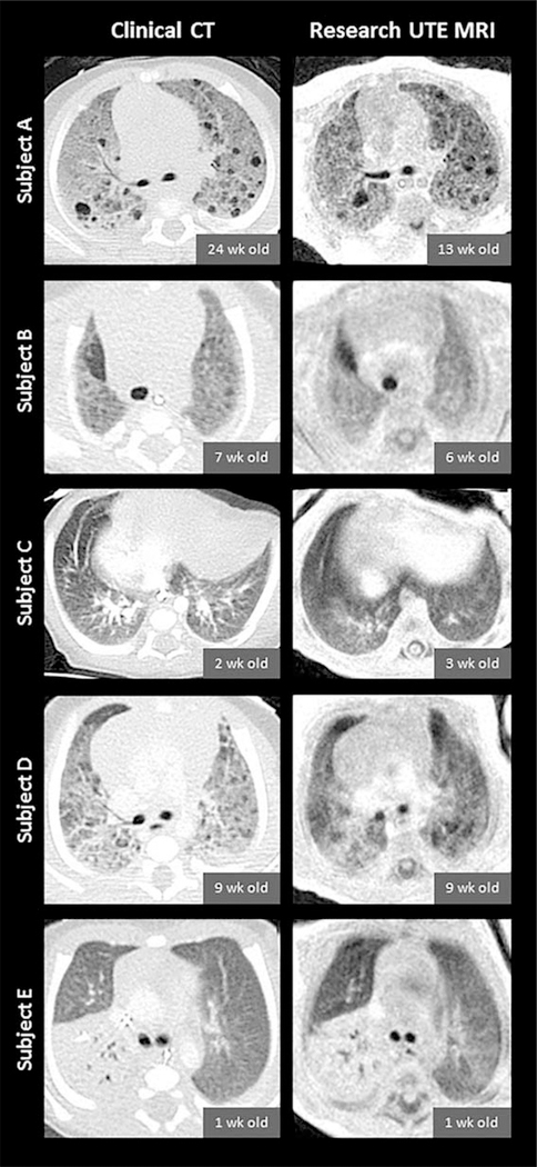
FIGURE 1:
Slice-matched images comparing clinical CT scans (left column) and research UTE MRI scans (right column) in five diseased subjects (from top to bottom: Subjects A through E). CT and MRI exams were typically performed at different timepoints for each individual subject; chronological ages at each subject’s CT and MRI exams are provided. CT images have an in-plane isotropic pixel resolution of 0.29–0.39mm with a slice thickness of 2.0–3.8mm, and UTE images have an in-plane isotropic pixel resolution of 0.70mm with a reconstructed slice thickness of 2.8mm (reconstructed at 4× thicker than the nominal UTE z-resolution of 0.70mm in order to optimize slice-matching). Note that contrast was administered for the clinical CTs of Subjects C, D, and E, and that the spinal-column appearance is influenced by components with different T1 and , similar to lung.

