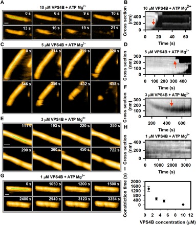
Fig. 3. Effect of VPS4 concentration on CHMP2A-CHMP3 tube remodeling in presence of ATP Mg2+.

(A) Clips of HS-AFM images captured at 1 frame/s, showing a rapid disassembly of tube, treated with 10 μM VPS4B. (B) Kymograph taken from (A), showing the time and position of first constriction and/or disassembly (marked by red arrow). (C) Same as (A) but for 5 μM VPS4B-treated tubes. Frame rate, 1 frame/s. (D) Same as (B) but derived from (C). (E and F) Same as (C) and (D), respectively, but for 3 μM VPS4B-treated tubes. One can see that, at this concentration, the tube goes through constriction but not disassembly. (G and H) Same as (C) and (D), respectively, but for 1 μM VPS4B-treated tubes. Images in (G) show that at, 1 μM VPS4B concentration (with ATP Mg2+), the tube undergoes a partial constriction after a significant amount of time. Frame rate, 0.33 frame/s. (I) Effect of VPS4B concentration on the cleavage of CHMP2A-CHMP3 tubes. The cleavage time is considered from the moment of addition of 200 μM ATP Mg2+ until the first cleavage occurs (red arrows in the kymograph). For four different concentrations of VPS4B, more than 20 tubes were investigated. Scale bars, 100 nm.
