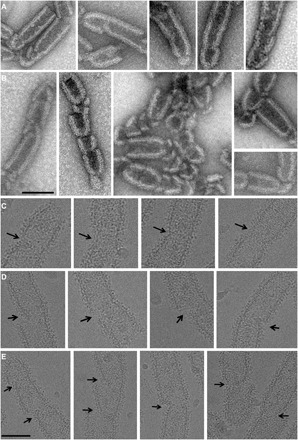
Fig. 4. EM imaging of CHMP2A-CHMP3 tube remodeling by VPS4B.

(A and B) Negative-stain images of CHMP2A-CHMP3 tubes incubated with 5 μM VPS4B and 200 μM ATP Mg2+, indicating (A) start sites of cleavage and (B) the generation of dome-like end caps. Scale bar, 100 nm. (C to E) Cryo-EM images of CHMP2A-CHMP3 tubes incubated with 5 μM VPS4B and 200 μM ATP Mg2+. (C) Images of early stages of constriction sites, (D) asymmetric constriction start sites, and (E) the generation of dome-like end caps. Scale bar, 100 nm.
