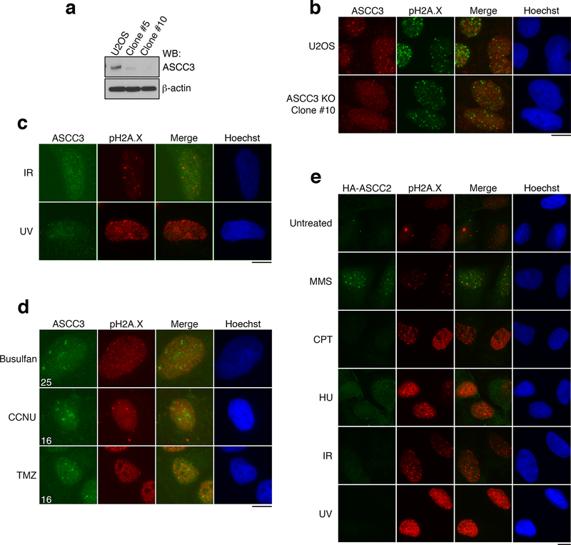
Extended Data Figure 1. The ASCC complex forms foci upon alkylation damage.
(a) ASCC3 KO cells were generated using CRISPR/Cas9 technology. Lysates were analyzed by Western blotting (n=2 independent experiments). Clone #10 was verified to be a knockout by deep sequencing. (b) Images of U2OS parental cells or ASCC3-KO cells after MMS (n=3 biological replicates). (c) Immunofluorescence of U2OS cells after exposure to γ-irradiation (IR; 5 Gy) or UV (25 J/m2) (n=3 biological replicates). (d) Images of U2OS cells after treatment with the alkylating agents busulfan (4 mM), 1-(2-chloroethyl)-3-cyclohexyl-1-nitrosourea (CCNU; 100 μM), or temozolomide (TMZ; 1.0 mM) (n=2 biological replicates). Numbers indicate the mean percent of cells expressing five or more foci. (e) Immunofluorescence of HA-ASCC2 expressing cells after exposure to the indicated damaging agents (n=3 biological replicates). Scale bars, 10 μm. For gel source data, see Supplementary Figure 1.

