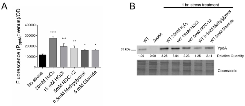
Figure 3: Oxidative and electrophilic stresses increase YpdA expression.

(A) ypdA promoter activity in S. aureus upon exposure to oxidizing and electrophilic agents. Overnight cultures were diluted in fresh TSB and allowed to grow to OD600=0.4, stressor agents were then added. Venus YFP fluorescence (515 nm excitation, 533nm emission) generated from the ypdA promoter activity was measured at 1-hour after the addition of stressor agent. Fluorescence data were normalized by optical densities. Values represent the mean and standard deviation of three biological replicates that were conducted in triplicate. Statistical significance between stress and no stress conditions was calculated using 1-way ANOVA Dunnett’s multiple comparisons test (* P< 0.05; ** P< 0.01; *** P< 0.001; **** P< 0.0001).
(B) YpdA protein levels in S. aureus increase with stress. Overnight cultures were diluted in fresh TSB and allowed to reach OD600=0.4 followed by the addition of stressor agents and cultures were further incubated for 1 hr. Fifteen ug of whole cell lysate were immunoblotted and probed with mouse anti-YpdA serum diluted 1:1000 followed by secondary antibody and substrate. Relative quantification of YpdA were analyzed by ImageJ; data were shown below the blot with WT set at 1.00. A parallel run gel stained with Coomassie is shown in the bottom panel to demonstrate comparable loading. Experiments were performed at least three times; a representative experiment is displayed.
