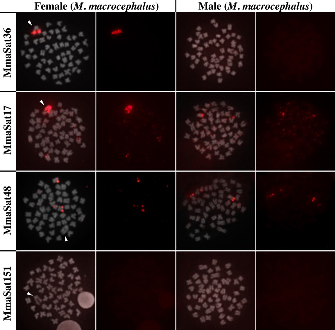
Figure 1.
Examples of the chromosomal distribution patterns of the mapped satDNAs in M. macrocephalus; those clustered solely on the W chromosome (MmaSat36); those clustered on the W chromosome and some autosomes (MmaSat17); those clustered on the autosomes (MmaSat48); and those that are nonclustered (MmaSat151). Each cell is shown with the satDNA FISH signal (red) merged with that of DAPI (left) and satDNA FISH (right). Arrowheads indicate the W chromosome. Bar = 10 μm.

