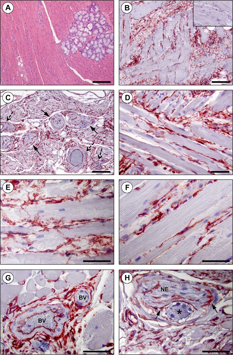
Figure 2.
Immunohistochemical localization of telocytes (TCs)/CD34+ stromal cells in the interstitium of the human tongue striated muscle. (A) Hematoxylin and eosin staining demonstrating the normal appearance of the tongue muscle. (B–H) CD34 immunohistochemistry with hematoxylin counterstain. (B,C) A diffuse CD34+ reticular network is evident in the perimysium encasing skeletal muscle bundles (B) and around intramuscular vessels (dashed arrows) and nerves (arrows) (C). Inset: negative control. (D–F) The endomysium is populated by a dense meshwork of CD34+ TCs projecting long and moniliform telopodes in close relationship with skeletal muscle fibers. (G) CD34+ TCs intimately encircle intramuscular arterioles (BV, blood vessels). (H) TCs form an outer sheath (arrows) for intramuscular nerves (NE) and ganglia (asterisk). Scale bar: 200 µm (A), 100 µm (B,C), 50 µm (D–H).

