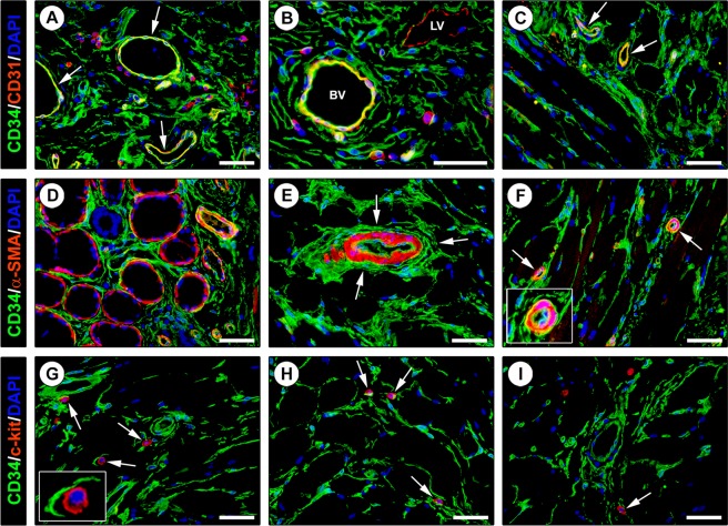
Figure 3.
Double immunofluorescence staining of human tongue tissue sections. (A–C) CD34 (green) and CD31 (red) immunostaining with 4′,6-diamidino-2-phenylindole (DAPI; blue) counterstain for nuclei. Telocytes (TCs)/CD34+ stromal cells in the tongue lamina propria (A,B) and skeletal muscle (C) lack CD31 immunoreactivity. The endothelial cells of blood vessels (arrows) are CD34+/CD31+ (A,C). At variance with double stained blood vessels (BV), the endothelium of lymphatic vessels (LV) is CD34−/CD31+ (B). Some leukocytes are also CD31+ (A,B). (D–F) CD34 (green) and α-smooth muscle actin (α-SMA; red) immunofluorescence labeling with DAPI counterstain. CD34+ TCs do not coexpress α-SMA. (D) CD34+ TCs envelop secretory salivary gland units outside of α-SMA + myoepithelial cells. (E,F) CD34+ TCs externally encircle the vascular smooth muscle cell layer of arterioles (arrows; higher magnification in the inset). (G–I) CD34 (green) and c-kit/CD117 (red) immunofluorescence with DAPI counterstain. The CD34+ TC meshwork is immunophenotypically negative for the c-kit/CD117 marker. c-kit/CD117 is detectable only in oval/round-shaped mast cells (arrows) often in close relationship with CD34+ TC processes (higher magnification in the inset). Scale bar: 50 µm (A–I).

