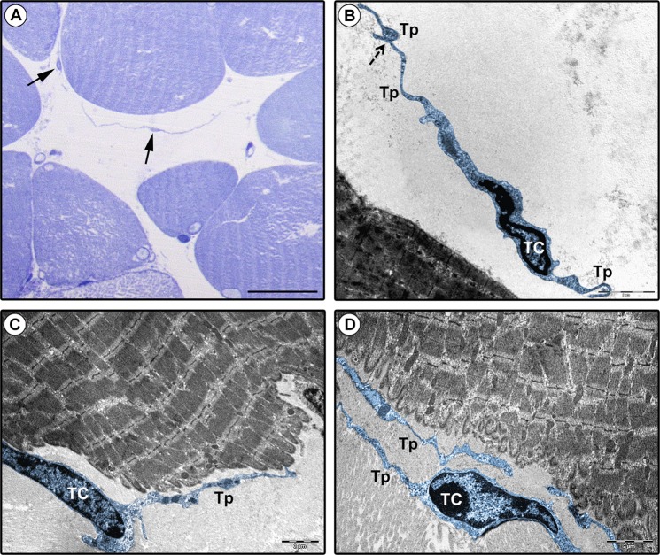
Figure 5.
Ultrastructural identification of telocytes (TCs) in human tongue muscle interstitium. (A) Semithin sections stained with toluidine blue and observed by light microscopy. Spindle-shaped cells with very long and thin moniliform cytoplasmic processes (arrows) are observed in the stroma surrounding skeletal muscle fibers. (B–D) Tongue muscle ultrathin sections stained with UranyLess and bismuth subnitrate solutions and observed by transmission electron microscopy. TCs (digitally colored in blue) are ultrastructurally identifiable as interstitial cells with telopodes (Tp), namely cytoplasmic prolongations with a moniliform silhouette due to the alternation of thin segments (podomers) and expanded portions (podoms). TCs may display a spindle-shaped, oval or piriform cell body mostly occupied by the nucleus. Telopodes are often interconnecting (B, dashed arrow) and in close relationship with skeletal muscle fibers (C,D). Scale bar: 25 µm (A), 2 µm (B–D).

