
Figure 1.
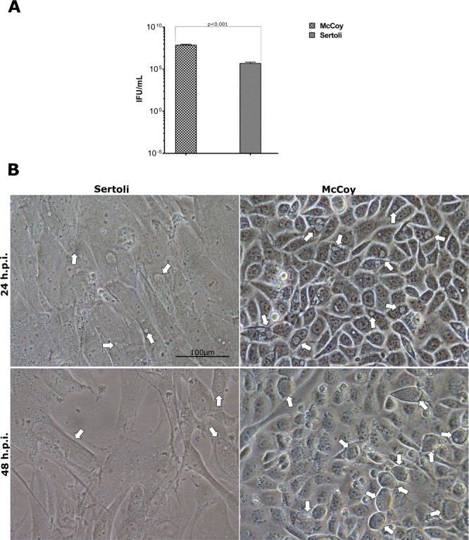
Comparison of C. trachomatis infectivity and morphological phenotype in primary human Sertoli and McCoy cells. (A) Yield of C. trachomatis D/UW-3/CX infection of primary human Sertoli and McCoy cells at a MOI = 1.0, expressed as means ± SD of four replicates from two independent experiments; (B) Phase-contrast micrographs of C. trachomatis inclusions in primary human Sertoli and McCoy cells at 24 and 48 hours post infection. Representative images of ten microscope fields are shown. Arrows point to chlamydial inclusions.