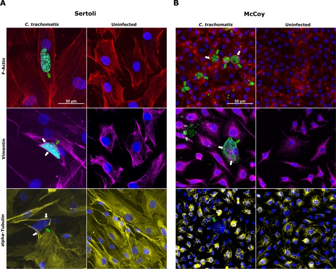
Figure 4.
Confocal analysis of cell cytoskeleton in primary human Sertoli and McCoy cells infected by C. trachomatis. Laser scanning confocal micrographs of F-actin microfilaments, Vimentin-based Intermediate Filaments and α-tubulin microtubules in primary human Sertoli (A) and McCoy (B) cells infected with C. trachomatis. Representative images of ten chlamydial inclusions are shown (100X magnification). White arrows point to assemblies of cytoskeleton fibres lining chlamydial inclusions. Green arrows point to C. trachomatis inclusions.

