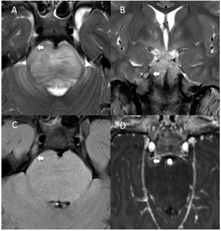
Figure 2.
Another MRI diagnosis of direct involvement of CN V by DIPG in a 7-years-old boy. TSE T2 axial (A) and coronal (B), axial FLAIR (C), and T1 post-contrast para-axial (D) reformatted MRI images show thickening and abnormal signal intensity involving the root entry zone and the cisternal course of the right V cranial nerve, contiguous with the tumor, (A–C, white arrows). Findings are consistent with direct right V cranial nerve involvement by the tumor. There is associated contrast enhancement (D, white arrow).

