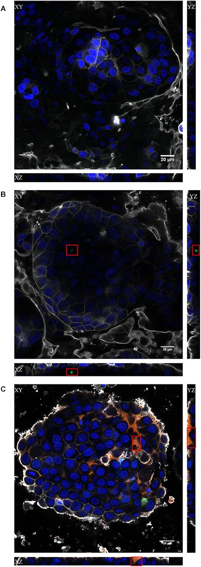
FIGURE 4.

Confocal images of the x, y, and z scans of the in vitro Caco-2/HT29/Raji-B lymphocytes co-culture model. The results obtained with the non-treated (A), bacteriophage-treated (B) and PPE (C) treated cultures at 2 h are shown. Cell nuclei were stained with Hoechst 33242 (blue), bacteriophages with SYBR gold (green), and liposomes with Vybrant Dil (red). The plasma membrane was stained with CellMask DeepRed (gray). Red squares indicate the regions where the stained phages (B) or PPE (C) were visualized. Scale bars, 20 μm.
