
FIGURE 5.
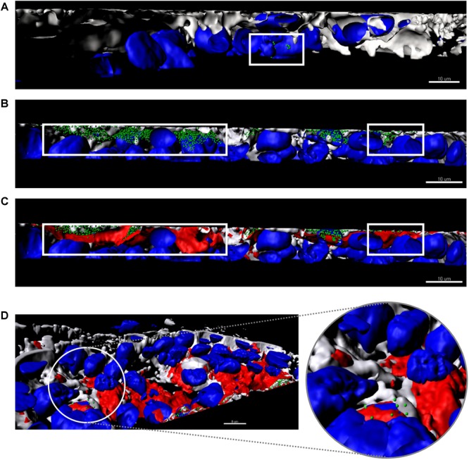
Three-dimensional images of a cross-section of the intestinal barrier model based on a Caco-2/HT29/Raji-B lymphocytes co-culture treated with bacteriophages (A) and PPE (B–D). Cell nuclei were stained with Hoechst 33242 (blue), bacteriophages with SYBR gold (green), and liposomes with Vybrant Dil (red). The plasma membrane was stained with CellMask DeepRed (gray). (A) Non-encapsulated phages (green) are seen inside the cells. (B,C) Stained PPE in different phases. (B) Only encapsulated phages (green) are shown inside the cells (gray, membrane and blue, cell nuclei). (C) Merging of all labels with the liposomes (red) covering the encapsulated phages detailed in (B). The white square indicates the non-encapsulated phages (A), the encapsulated phages (B), and liposome capsule (C). (D) A detail of the basolateral side of the intestinal barrier (sited in the upper side of the image) shows the presence of liposomes (red) inside the cells. The enlarged image shows phages released from the liposomes. Scale bars, 8 μm.