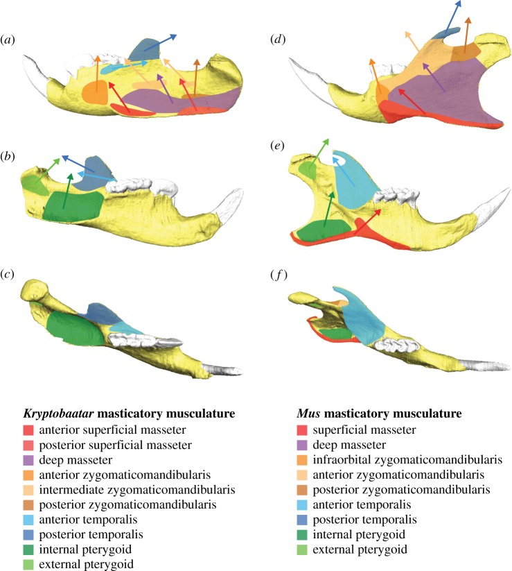
Figure 3.
Muscle attachment sites and vectors on the hemimandibles of (a–c) Kryptobaatar and (d–f) Mus. (a) and (d) in lateral view, (b) and (e) in medial view, (c) and (f) in occlusal/dorsal view. Attachment sites are based on [35] and [41]. Muscle vector orientations were calculated during finite-element model construction in HyperMesh software (see Material and methods).

