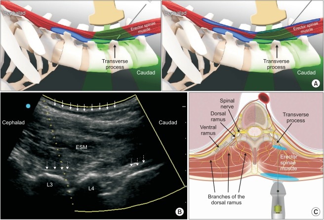
Fig. 1.
(A) In the conventional erector spinae plane (ESP) block, local anesthetic is injected into the musculofascial plane deep to the erector spinae muscle (left panel). In the modified dual-injection ESP block, a second injection of local anesthetic is made into the musculofascial plane superficial to the erector spinae muscle as a needle is withdrawn towards the skin (right panel) (Image used with permission from Twin Oaks Anesthesia Services, LLC). (B) Longitudinal parasagittal ultrasonographic view of the erector spinae muscle (ESM) overlying the articular and transverse processes of the L3 and L4 vertebrae. A dark hypoechoic layer of local anesthetic (solid arrows) can be seen spreading superficial to the hyperechoic posterior investing fascia of ESM. Another layer of local anesthetic (dotted arrows) is visible deep to ESM and superficial to the hyperechoic bony surfaces of the vertebral column. (C) The branches of the dorsal rami of spinal nerves innervate the vertebrae and paraspinal tissues. By depositing local anesthetic (blue shaded areas) into two separate fascial planes superficial and deep to the erector spinae muscle, the branches of the dorsal rami are blocked at both proximal and distal locations (Image adapted and used with permission from Maria Fernanda Rojas Gomez).

