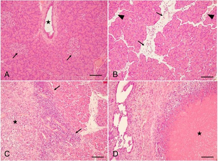
Fig. 3.
Histological findings in normal and pancreatitis-induced pigs. All tissue sections were stained with hematoxylin–eosin. a Normal pancreas showing an intralobular pancreatic duct (star), acini and endocrine islets (arrows). b Mild disease with scattered inflammatory cells and interlobular edema (arrows) as well as interacinar edema (arrowheads). In this example, the total histopathological pancreatitis score was 3. c Moderate disease with dense inflammatory infiltrates intralobular (arrows) as well as focal tissue necrosis (star). In this example, the total histopathological pancreatitis score was 6. d Severe disease with extensive tissue necrosis (star) and dense inflammatory cells around the necrotic area. In this example, the total histopathological pancreatitis score was 11. All bars equal 50 μm

