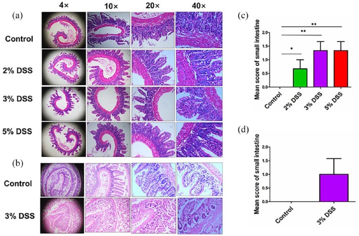
Figure 3.
Histological observation of DSS-induced AEE. (a) Representative schematic of the H&E staining in small intestine tissues in mice treated by different concentrations of DSS in cohort 1. (b) Representative schematic of the H&E staining in small intestine tissue in mice treated by 3% DSS in cohort 2. (c) Comparison of histopathological scores of small intestine in mice treated by different concentrations of DSS in cohort 1. (d) Comparison of histopathological scores of small intestine in mice treated by 3% DSS in cohort 2. *P < 0.05; **P < 0.01.

