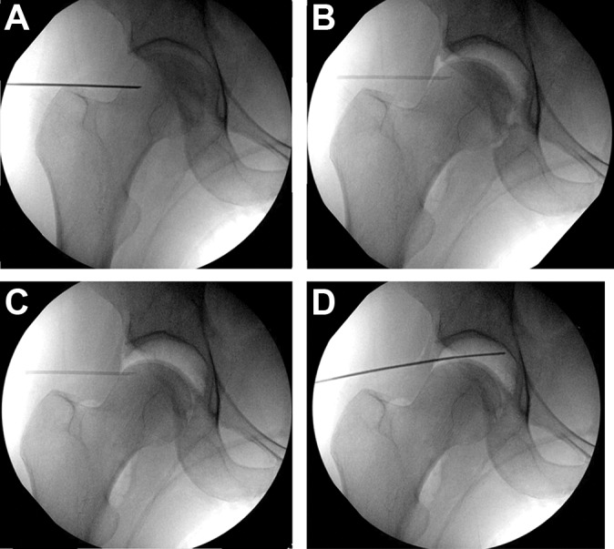
Figure 1.

(A) A 17-gauge spinal needle is used to penetrate the hip joint capsule anteriorly, away from the labrum and weightbearing articular surface to reduce the risk of iatrogenic injuries. (B) Afterward, 25 mL of air is injected through the spinal needle. The subsequent air arthrogram confirms the intra-articular position, breaks the suction seal of the hip joint, and aids in distraction. (C) After 10 turns of fine traction is applied, the hip joint easily distracts, allowing a safe space for instrumentation without damage to the labrum or articular cartilage. (D) Redirecting the spinal needle allows a pathway for instrumentation into the joint over a guide wire, with continued fluoroscopic guidance until the arthroscope is inserted and the remainder of the procedure can be directly visualized.
