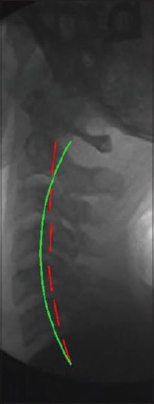
Figure 2.

Postcervical lordosis adjustment radiograph. Lateral cervical images were analyzed using PostureRay® EMR software. Postradiographs show the patients with the Cervical Denneroll™ Spinal Orthotic applied as a fulcrum to the cervical spine yielding an increase in cervical lordosis. Each image was analyzed for cervical lordosis measurements using the Harrison posterior tangent method. Red indicates the posterior aspect of the cervical vertebrae from C2 to C7. Green indicates ideal cervical lordosis from C2 to C7
