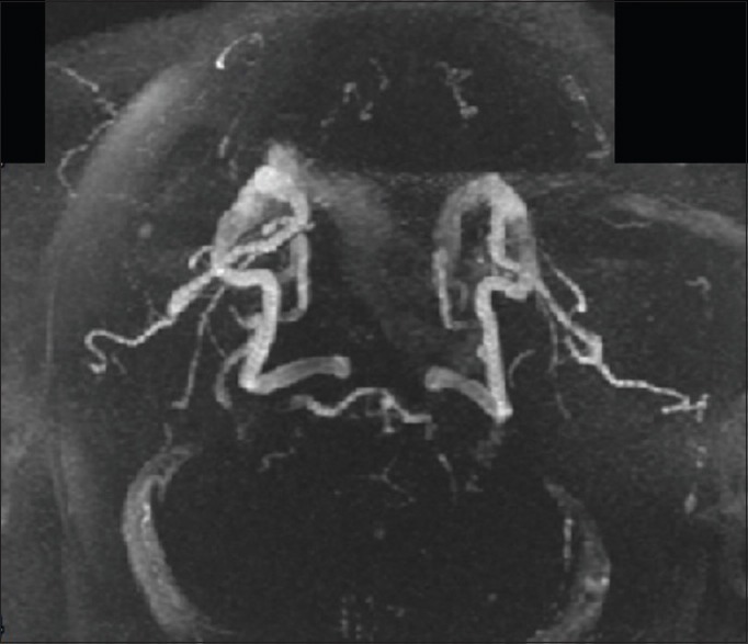
Figure 5.

Postcervical lordosis adjustment magnetic resonance angiogram. Brain magnetic resonance angiogram images were analyzed using FIJI/ImageJ. Each image set was converted to an 8-bit TIF format, and appropriate pixel thresholds were set for each image pair using the threshold tool. White indicates blood in cerebral vasculature
