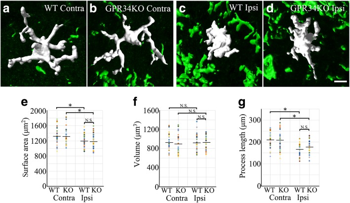
Fig. 3.
Microglial morphology is not affected by GPR34 deficiency. a–d Representative 3D reconstructions of microglia in the contralateral (Contra) and ipsilateral (Ipsi) dorsal horn in WT and GPR34-deficient mice. Scale bar = 10 μm. e–g Quantitative morphometric analysis of microglial surface area (e), volume (f), and process length (g) (n = 32 cells from four animals). Values are mean ± SEM. *p < 0.05 (one-way ANOVA with post-hoc Tukey’s test). There was no significant difference in any parameter between WT and GPR34-deficient mice

