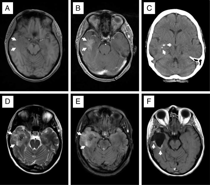
Fig. 1.
Neuroradiologic imaging pattern of primary brain amyloidoma. The T1-weighted MRI sequence shows a slightly hypointense right temporal mass (arrows in a), an ill-defined, moderate gadolinium enhancement (arrows in b), both compatible with malignant glioma. The CT displays small focal calcifications (arrows in c). The corresponding T2- and FLAIR MRI sequences displayed a hyperintense signal extending beyond the margins of the contrast enhancement indicating peritumoral edema or an infiltrating part of the mass (arrows in d and e). Almost 2 years following surgical removal of the PBA, the T1-weighted MRI sequence shows contrast enhancement at the resection margins (arrows in f) compatible with residual amyloidoma that remained unchanged during the 8-year follow-up

