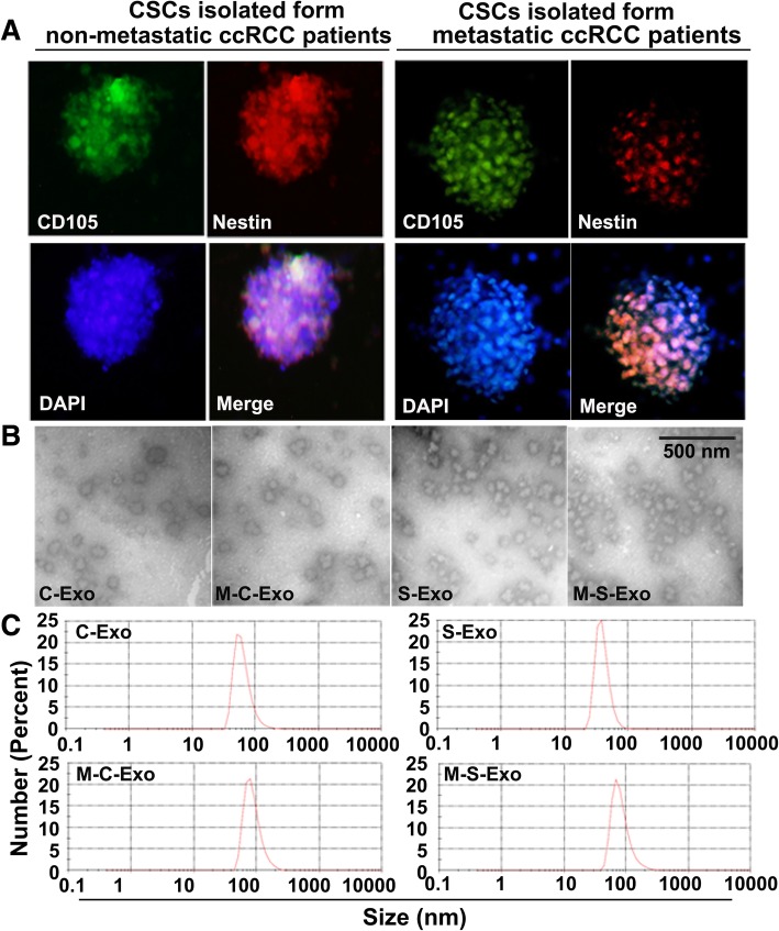
Fig. 1.
Characterization and measuring of exosomes isolated from CCRCC patients. a Immunofluorescent image showing the positive expression of CCRCC stem cells (CSCs) markers CD105 and Nestin. b Representative transmission electron micrographic images of four types of exosomes from CCRCC patients of different pathological stages and cell origins. C-Exo: exosomes isolated from cancer cells of non-metastatic CCRCC patients; M-C-Exo: exosomes isolated from cancer cells of lung-metastatic CCRCC patients; S-Exo: exosomes isolated from cancer stem cells (CSCs) of non-metastatic CCRCC patients; M-C-Exo: exosomes isolated from CSCs of lung-metastatic CCRCC patients. c Nanoparticle tracking showing the diameter of the particles

