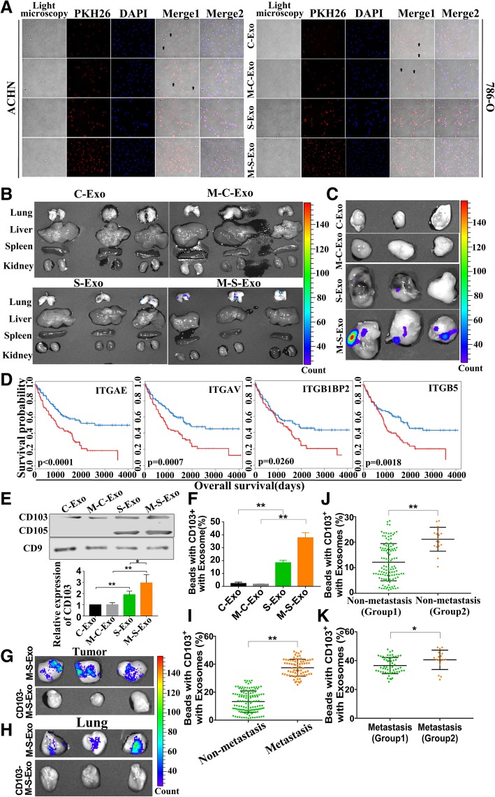
Fig. 6.
CD103+ CSCs exosomes contribute to the cancer cell organotropism. a Relative ability of varying types of exosomes to fuse with CCRCC cells, as assessed by fluorescent labelling with PKH26 membrane dye in ACHN and 786-O cells. Arrows indicate no fusion. b Comparative distributions of PKH26-labeled exosomes of different types in the main organs of nude mice. n = 6 mice per treatment group. c Comparative distributions of PKH26-labeled exosomes of varying types in the resected tumor xenografts. n = 6 mice per treatment group. d Correlation of expression levels of four integrins (ITGAE/CD103, ITGAV, ITGB1BP2, and ITGB5) with the overall survival rate of CCRCC patients. e The CD103 protein levels in C-Exo, M-C-Exo, S-Exo and M-C-Exo. n = 5, *P < 0.05, **P < 0.01 relative to the controls. f The ratio of CD103+ exosomes over total exosomes of four different types determined by flow cytometry quantification. g & h Removal of CD103+ exosomes (CD103− M-S-Exo) rendered a loss of the tumorigenesis and aggregation of M-S-Exo in tumor and lung. n = 6 mice per treatment group. i The ratio of CD103+ exosomes over total exosomes in the blood samples from non-metastatic and metastatic CCRCC patients, determined by flow cytometry quantification. j & k Before and after cancer metastasis in 17 CCRCC patients, the ratio of CD103+ exosomes in the blood samples were detected and compared with other patients without and with metastasis respectively. Group 2 represents the exosomes isolated from 17 CCRCC patients

