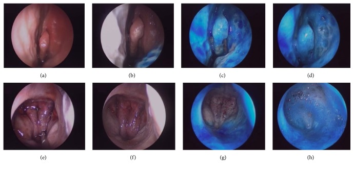
Figure 2.
Representative endoscopic images of the left nostril during various stages of blue dye treatment. At the middle turbinate: nasal mucosa prior to treatment (a) and following nasal spray (b), MAD nasal (c), and Spray-sol (d) applications; at the nasopharynx: nasal mucosa prior to treatment (e) and following nasal spray (f), MAD nasal (g), and Spray-sol (h) applications.

