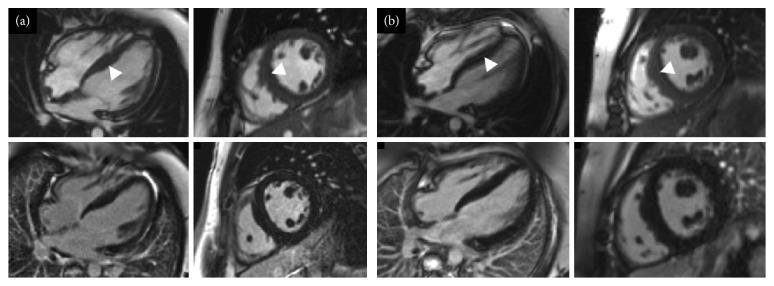
Figure 3.
Short-axis and four chamber cine images from the initial cardiovascular magnetic resonance scans of II:2 (a) and II:3 (b) from family A. Top row for each set of four images shows cine images and the bottom row late gadolinium enhancement images. (a, b) Mild asymmetric left ventricular hypertrophy (maximum wall thickness: (a) 13 mm; (b) 14 mm). No late gadolinium enhancement is demonstrated.

