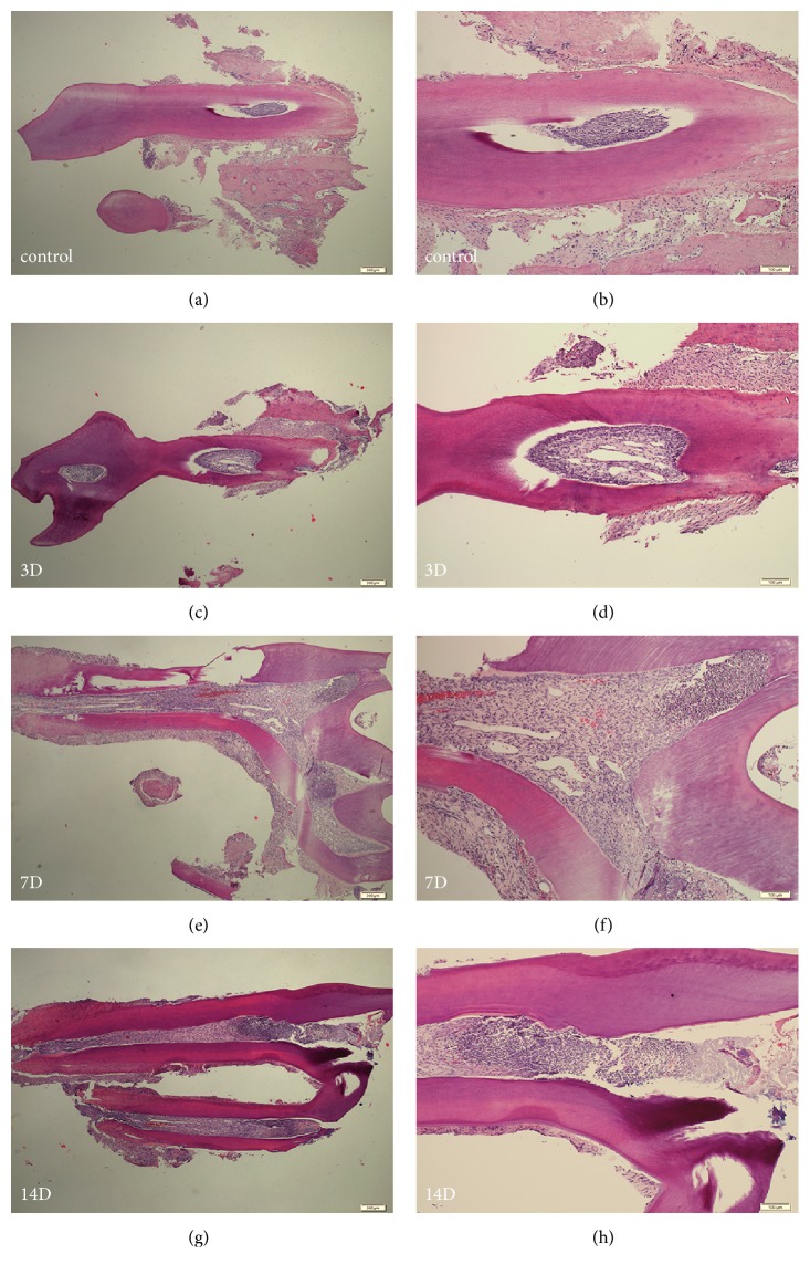
Figure 2.
Hematoxylin-eosin staining of sagittal sections of left first maxillary molars. (a) Sham-operated rat at 4×magnification. (b) Sham-operated rat at 10×magnification. (c, e, g) The cavities were made, and acute inflamed dental pulps at 3, 7, and 14 days are shown at 4×magnification. (d, f, h) The cavities were made, and acute inflamed dental pulps at 3, 7, and 14 days are shown at 10×magnification.

