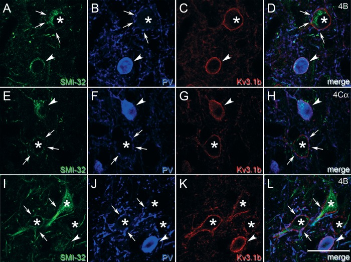
Figure 7.
High magnification images compare cell membrane staining of Kv3.1b in PV-ir and non-PV-ir neurons. Single z plane images from sampled regions in layer 4B (A–D, I–L) and 4Cα (E–H) of animal M1. These regions contained neurons that were both Kv3.1b-ir and PV-ir (arrowheads) and neurons that were Kv3.1b-ir but not PV-ir (asterisks). Although the Kv3.1b-ir, non-PV-ir neurons appeared to be surrounded by perisomatic baskets of PV-ir terminals, some of which are indicated by arrows, there was also continuous membrane Kv3.1b staining apparent in the intervening regions. At least 2 of these Kv3.1b-ir, non-PV-ir neurons—asterisk in A–D and uppermost asterisk in I–L—had the shape of pyramidal cells and were SMI-32-ir. Scale bar: 25 μm.

