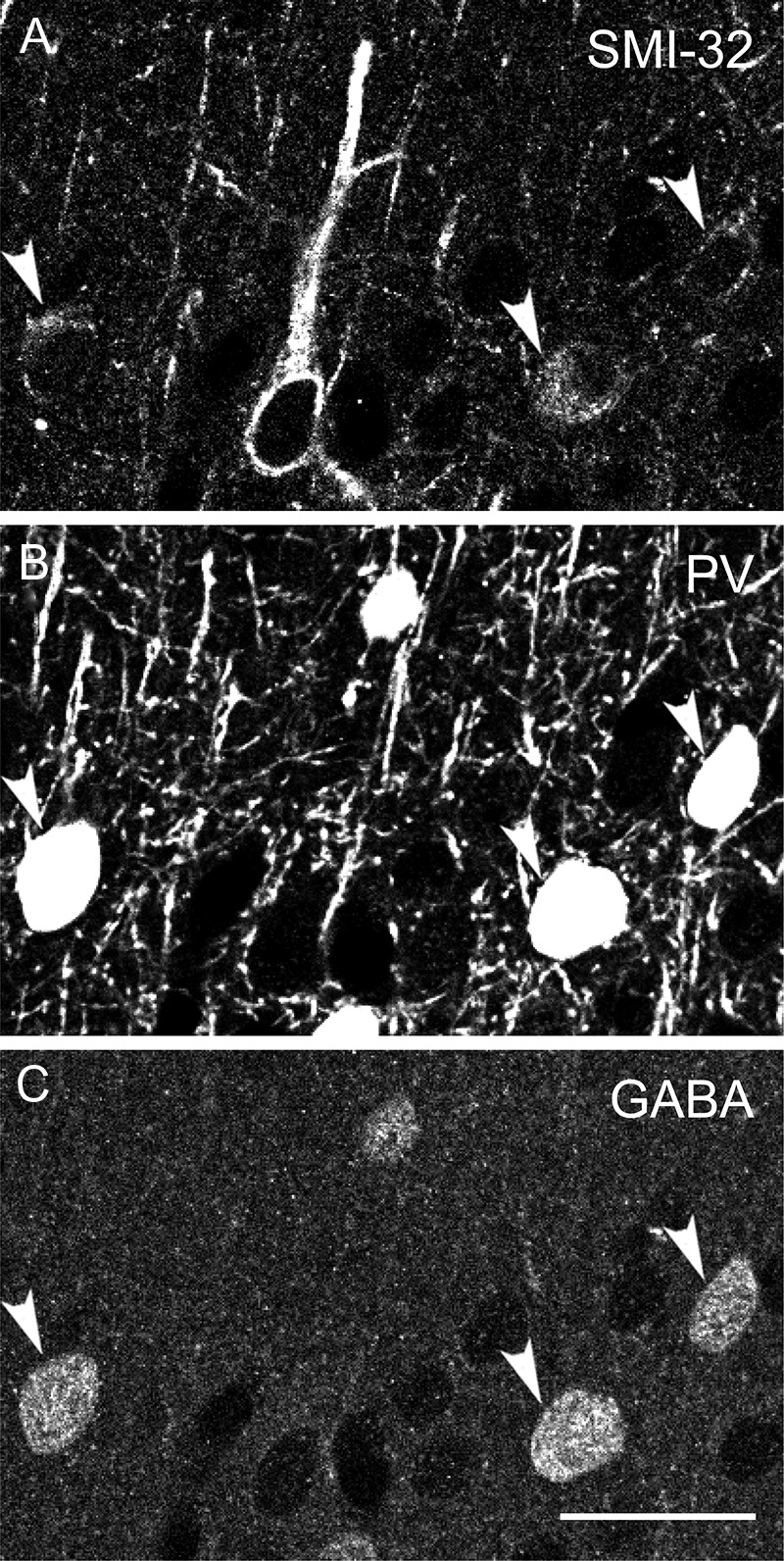
Figure 8.

PV-, GABA-ir, and SMI-32-ir neurons. Confocal micrographs of tissue from animal M3 triple immunofluorescence labeled with SMI-32 (A), anti-PV (B), and anti-GABA (C). Arrowheads indicate examples of SMI-32-ir, PV-ir, GABA-ir neurons. Scale bar in C (refers to all panels): 25 μm.
