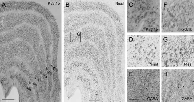
Figure 9.
Kv3.1b in the LGN. (A): Micrograph of coronal section from dorsal LGN of monkey M5 showing pattern of Kv3.1b expression. (B): Adjacent Nissl stained section. (C–E): Enlarged view of Kv3.1b (C), Nissl (D), and GABA (E) staining in magnocellular layer 1 shows Kv3.1b-ir neurons are prevalent and may include the entire excitatory population. Location of enlarged region identified in (B). (F–H): Enlarged view of Kv3.1b (F), Nissl (G), and GABA (H) staining in parvocellular layer 4. Location of enlarged region identified in (B). Scale bar in (A) (refers to A and B): 500 μm. Scale bar in H (refers to C and H): 100 μm.

