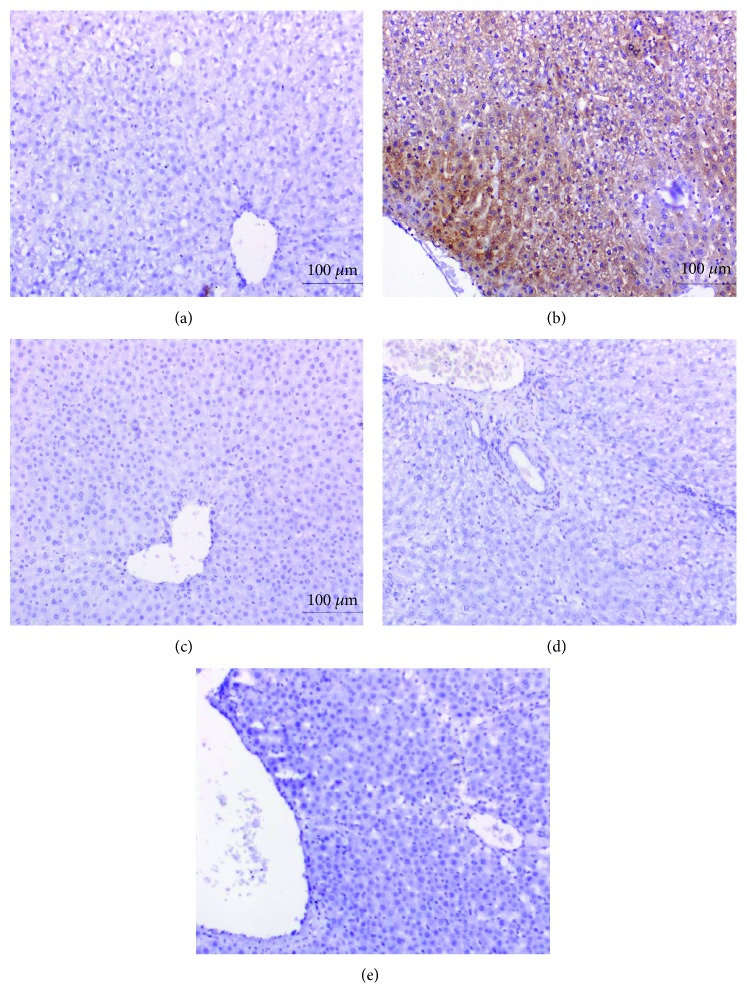
Figure 5.
Immunohistochemically stained liver sections for Bax expression detection. (a) Negative or very weak immunohistochemical reaction in normal control. (b) Evoked a strong positive immunohistochemical reaction marked by a dense brown colour in the cytoplasm of hepatocytes in APAP-administered rats. (c-e) Profound suppression of Bax expression in APAP-administered rats treated with the navel orange peel extract (c), naringin (d), and naringenin (e) (×200).

