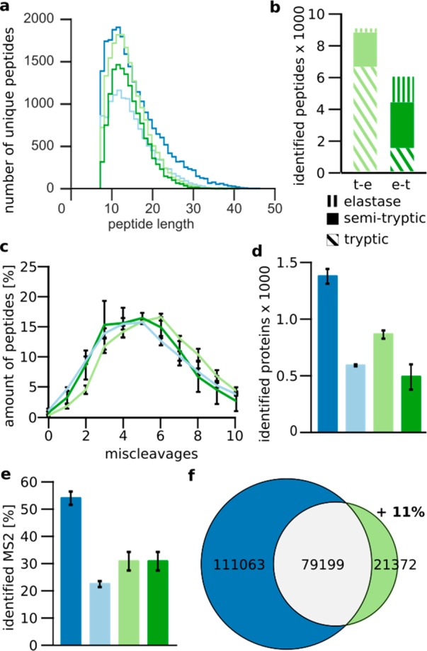
Figure 4.

Impact of elastase when following trypsin in a sequential digest in S. pombe cell lysate. (a) Peptide-length distribution. (b) Number of tryptic, semitryptic, and elastase peptides identified in both the trypsin–elastase (light green) and elastase–trypsin digests (dark green). (c) Distribution of miscleavages for elastase, trypsin–elastase, and elastase–trypsin digests. (d) Number of identified proteins. (e) Percentage of identified MS2. (f) Overlap of observed residues by trypsin and sequential trypsin–elastase digestion. Data shown are the means ± SD of duplicate injections from three independent digestions. Trypsin, dark blue; elastase, light blue; sequential trypsin–elastase, light green; sequential elastase–trypsin digest, dark green.
