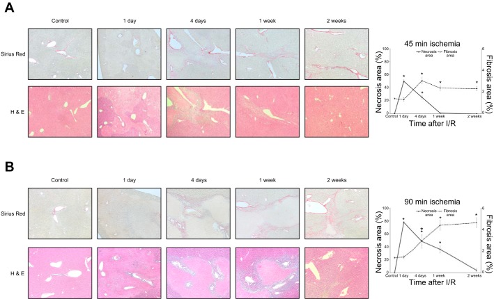
Fig. 1.
Development of liver fibrosis after ischemia-reperfusion (I/R) injury. A: liver sections from mice undergoing 45-min ischemia followed by reperfusion were analyzed. Sirius red staining of liver sections shows increased collagen deposition 4 days after I/R injury and maintaining a similar level of fibrosis up to 2 wk after I/R. Hematoxylin-eosin staining shows significant liver injury 1 day after reperfusion with repair and resolution of injury to nearly normal liver architecture by 1 wk after reperfusion. Original magnification is ×100. B: liver sections from mice undergoing 90-min ischemia followed by reperfusion were analyzed. Sirius red staining of liver sections shows collagen deposition within 4 days after I/R injury and increasing fibrosis during the time of liver repair (up to 2 wk after I/R). Hematoxylin-eosin staining shows massive liver injury 1 day after I/R with repair and resolution of the injury to nearly normal liver architecture by 2 wk after reperfusion. There was much more liver injury and liver fibrosis in mice undergoing 90 min of ischemia compared with those undergoing 45 min of ischemia. Original magnification is ×100. Quantification of necrosis area and fibrosis area shows an inverse relationship between necrotic area and extent of liver fibrosis. Data are means ± SE with n = 3 per group. *P < 0.05 compared with control.

