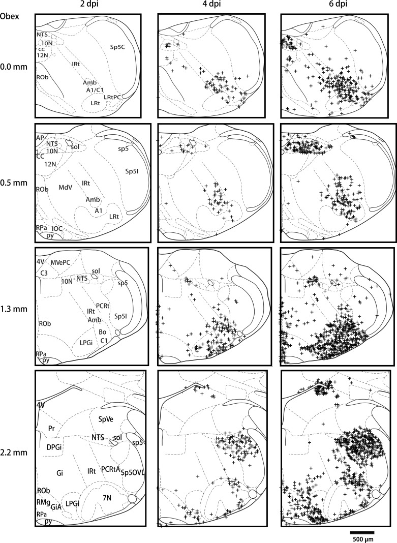
Fig. 3.
Schematic drawings showing typical nucleoprotein immunoreactivity (NP-IR) locations in coronal medullary sections at 0, 0.5, 1.3, 2.2 mm rostral to the obex found in 3 HK483 mice at 2, 4, and 6 days postinfection (dpi). NP-IR is not detectable until 4 dpi. NP-IR is detected in all of those levels, but it is relatively higher in the rostral than the caudal regions. This is more widespread in the LRt, 10N, LC, RPa, Amb, and LPGi from 4 to 6 dpi compared with the other nuclei.

