Figure 2.

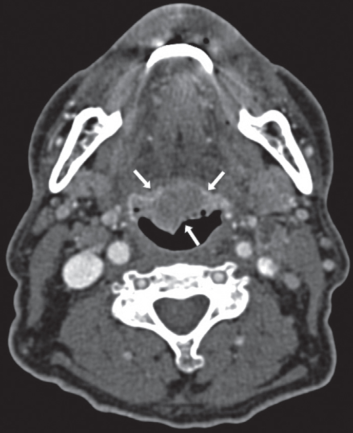
Axial slice of cervical CT showing a pathological mucous thickening of lingual tonsils, well delimited and of homogeneous density (arrows)

Axial slice of cervical CT showing a pathological mucous thickening of lingual tonsils, well delimited and of homogeneous density (arrows)