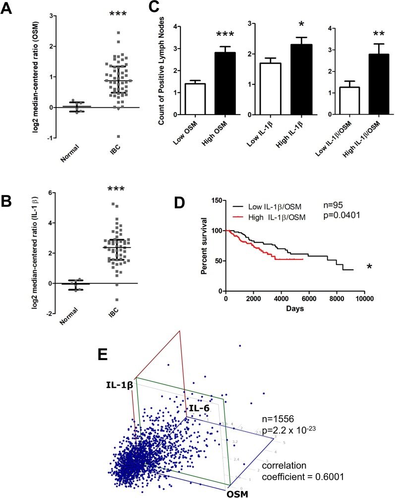
Figure 6. OSM and IL-1β expression is higher in invasive breast cancer compared to normal tissue and correlates with higher lymph node metastasis, decreased survival, and IL-6 levels.
(A) Using the FINAK dataset obtained from Oncomine™ we assessed stromal tissue expression of OSM and IL-1β. Stromal tissue expression of OSM is 5.9-fold higher in invasive breast cancer patients compared to normal patients. (B) Similarly, expression of IL-1β is 5.4-fold higher in the stromal tissue of invasive breast cancer patients compared to normal patients. (C) Using the Curtis dataset obtained from Oncomine™, we correlated OSM and IL-1β tissue expression levels to the number of lymph node metastases. Patients with high OSM (Left), high IL-1β (Center), and high co-expression of both OSM and IL-1β (Right) have significantly higher number of lymph node metastatic nodules compared to the respective low expression group. (D) High co-expression of both OSM and IL-1β leads to a decreased overall patient survival. Log-rank test (p = 0.0401). (E) Expression of OSM, IL-1β, and IL-6 were analyzed in a three-way correlation analysis with OSM on the x-axis, IL-6 on the y-axis, and IL-1β on the z-axis. There is significant correlation with a coefficient of 0.6001, with a p-value of 2.2 × 10⁻²³. Bar and scatter plot data expressed as mean ± SEM, and significance assessed by two-tailed student’s t-test. *p < 0.05, **p < 0.01, ***p < 0.001.

