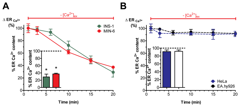
Fig. 2. Isolated pancreatic islets and β-cells have an atypical ER leak that is independent of SERCA activity.
(A,B) Percentage of ER Ca2+ leak, representing the maximal amount of Ca2+ released from the ER, under Ca2+-free conditions stimulated with the IP3-generating agonists (A) carbachol (100 μM) in MIN-6 (red values) and INS-1 cells (green values) together with the SERCA-inhibitor BHQ (15 μM) or (B) histamine (100 μM) in HeLa (blue values) and EA.hy926 cells (black values) at the indicated time points. Bar charts represent the percentage of ER Ca2+ content after 20 min of incubation under Ca2+-free conditions. The corresponding 1 min value was set to 100% (n ≥ 6). *p<0.05 using one-way ANOVA.

