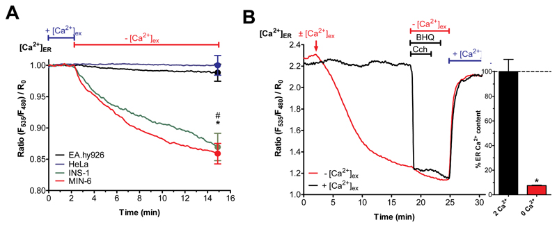
Fig. 3. Isolated pancreatic islets and β-cells have an atypical ER leak that is independent of SERCA activity.
(A) Curves reflect normalized [Ca2+]ER ratio signals over time measured with the genetically encoded Ca2+ probe D1ER in HeLa (blue curve), EA.hy926 (black curve), INS-1 (green curve) and MIN-6 (red curve) cells perfused with experimental buffer containing 2 mM Ca2+ for two min before switching to 0 mM Ca2+ and EGTA-containing buffer (n ≥ 6). (B) Representative curves for β-cells (depicted INS-1) reflect [Ca2+]ER ratio signals over time in the presence (2 mM Ca2+; black curve) or absence (0 mM Ca2+; red curve) of Ca2+ measured with D1ER. The ER Ca2+ store was depleted using carbachol (100 μM) and BHQ (15 μM) in Ca2+-free EGTA-buffered solution prior to addition of 2 mM external Ca2+ (n ≥ 6).

