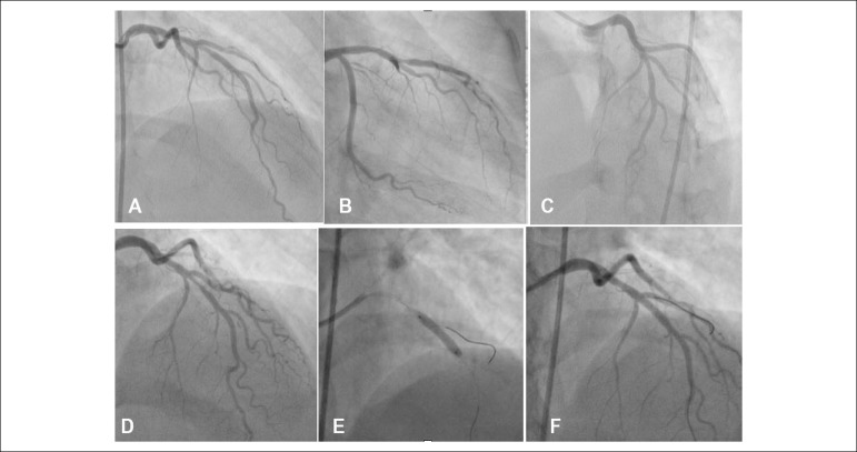
Figure 2.
Cardiac catheterization showing moderate stenosis in the middle third and moderate/severe segmental stenosis in the distal third of the anterior descending artery (ADA) in the cranial (A), right caudal (B) and left cranial (C) views. Restudy, after 3 months, showed a significant improvement of the obstruction in the distal third of the ADA, with moderate obstruction in the middle third, which was treated with the direct stenting technique (cranial view, images: D, E and F).

