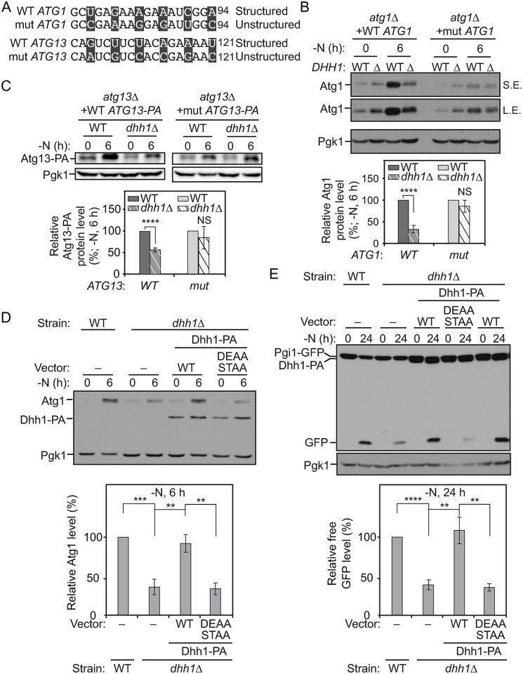
Fig 4. The structured regions in the ATG1 and ATG13 ORFs are necessary for the translational regulation by Dhh1 after nitrogen starvation.
(A) The mutations (“mut”) made in the structured regions of ATG1 and ATG13 ORFs are shown as indicated; mutated nucleotides are presented in black boxes. (B) WT Atg1 (XLY316), WT Atg1 dhh1Δ (XLY317), mutant Atg1 (XLY318), and mutant Atg1 dhh1Δ (XLY319) cells were grown in YPD to mid-log phase (-N, 0 h) and then shifted to SD-N for 6 h. Cell lysates were prepared, subjected to SDS-PAGE, and analyzed by western blot. The quantification of Atg1 protein level was conducted as indicated in Fig 2A. NS, not significant. ****p < 0.0001. (C) WT Atg13–PA (ZYY202), WT Atg13–PA dhh1Δ (ZYY203), mutant Atg13–PA (ZYY205), and mutant Atg13–PA dhh1Δ (ZYY206) cells were grown in YPD to mid-log phase (-N, 0 h) and then shifted to SD-N for 6 h. Cell lysates were prepared, subjected to SDS-PAGE, and analyzed by western blot. The quantification of Atg13–PA protein level was conducted as indicated in Fig 2A. The 5′-UTR and 3′-UTR of ATG13 in these strains were not changed. ****p < 0.0001. (D) The WT strain with empty vector (XLY329), the dhh1Δ strain with either empty vector (XLY331), or vectors expressing WT Dhh1–PA (XLY333) or Dhh1D195A,E196A,S226A,T228A–PA (DEAA STAA; XLY342) were grown in YPD to mid-log phase (-N, 0 h) and then shifted to SD-N for 6 h. Cell lysates were prepared, subjected to SDS-PAGE, and analyzed by western blot. The quantification of the Atg1 protein level was conducted as indicated in Fig 2A. **p < 0.01. ***p < 0.001. (E) The strains in (D) were grown in YPD to mid-log phase (-N, 0 h) and then shifted to SD-N for 24 h. Cell lysates were prepared and subjected to SDS-PAGE. The processing of Pgi1–GFP was analyzed by western blot. The quantification of free GFP level was conducted as indicated in Fig 1D. **p < 0.01. ****p < 0.0001. (See also S5 Fig; raw numerical values are shown in S1 Data). Atg, autophagy-related; DEAA STAA, Dhh1D195A,E196A,S226A,T228A; GFP, green fluorescent protein; L.E., long exposure; NS, not significant; ORF, open reading frame; PA, protein A; Pgi1, phosphoglucoisomerase 1; Pgk1, 3-phosphoglycerate kinase 1; SD-N, synthetic minimal medium lacking nitrogen; S.E., short exposure; WT, wild type; YPD, yeast extract–peptone–dextrose.

