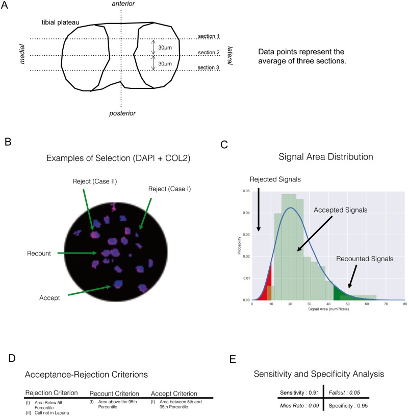
Fig 1. Computer assisted detection of chondrocytes and apoptotic chondrocytes in the histological sections of mouse knee joints.
A) Example for the classification of different DAPI signals for the quantification of chondrocytes. B) Area distribution of accepted vs. rejected signals. C) Criteria that define acceptance and rejection of a signal. D) Sensitivity and specificity of the automated detection under the given criteria. E) Histological sectioning and analysis.

