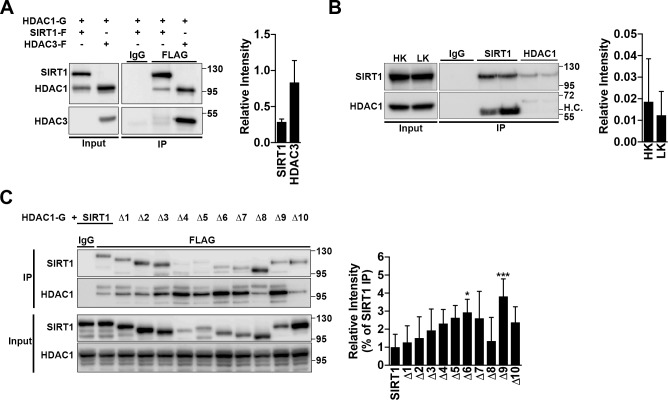
Fig 3. SIRT1 interacts with HDAC1.
A. Confirmation that SIRT1 and HDAC1 interact. HDAC1-GFP was co-expressed in a 1:1 ratio with either SIRT1-Flag or HDAC3-Flag into HEK293T cells. 48 h later, cells were lysed and immunoprecipitation (IP) was performed as indicated with either an IgG or Flag antibody. Membranes were probed with a GFP antibody to detect HDAC1 pulldown and then reprobed with a Flag antibody to check for proper pulldown of SIRT1 and HDAC3. Input represents 10% of whole cell lysate used for IP. Graph represents densitometry of HDAC1 pulldown from four independent experiments. B. Endogenous immunoprecipitation was performed on CGNs treated with HK/LK media for 6 h, with either an IgG, SIRT1, or HDAC1 antibody. H.C.: heavy chain. Graph represents densitometry from four independent experiments. C. Immunoprecipitation was performed as described in (A) with HDAC1-GFP co-expressed with flag-tagged SIRT1 or Δ1-Δ10. Pulldown was performed with either an IgG or Flag antibody. Membranes were initially probed with a GFP antibody and then stripped and reprobed for Flag. Graph represents densitometry (one-way ANOVA with Dunnett’s multiple comparisons posttest; *p<0.05, ***p<0.001) from five independent experiments and normalized to pulldown of full length SIRT1.

