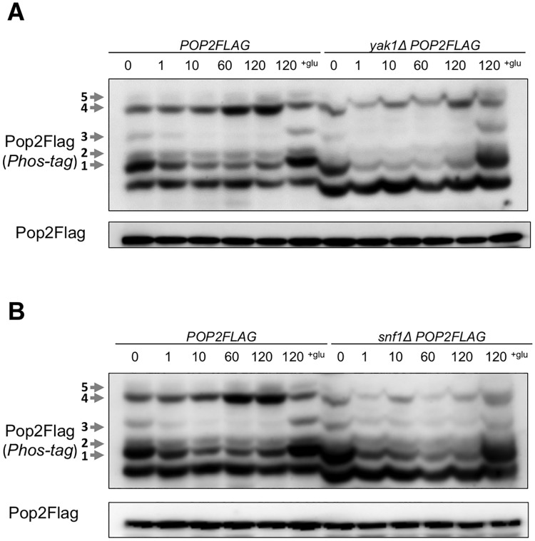
Fig 5. The phosphorylation of Pop2 in yak1Δ and snf1Δ.
(A) WT and snf1Δ cells harboring YCplac33-POP2FLAG (POP2FLAG, snf1ΔPOP2FLAG) were collected as in Fig 2A. Extracts prepared from each strain were run on phos-tag and conventional gels, then immunoblotted with anti-Flag antibody. Phosphorylated Pop2Flag is indicated with arrows and numbers according to the positions. Representative data are shown. (B) WT and yak1Δ cells harboring YCplac33-POP2FLAG (POP2FLAG, yak1ΔPOP2FLAG) were collected as in Fig 2A. Samples were analyzed as in (A). Representative data are shown.

