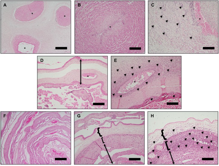
Fig 1. Representative placental histology.
(A&B) Normal umbilical cord. *indicates lumen of umbilical vessels. (C) Umbilical vein with heavy neutrophilic infiltrate (arrows point to aggregates of neutrophils) and early degeneration of smooth muscle cells diagnostic of severe fetal ACA. (D) Normal chorionic plate/villous parenchyma. Arrow indicates the chorionic plate, and * indicates lumen of a fetal chorionic plate vessel. (E) Chorionic plate with heavy neutrophilic infiltrate in the walls of a fetal chorionic plate vessel (severe fetal ACA) and neutrophils in the subchorionic fibrin layer (maternal ACA). Arrows point to aggregates of neutrophils. (F&G) Normal membrane roll. (H) Membrane roll with neutrophilic microabscess diagnostic of severe maternal ACA (arrows point to neutrophils). In G and H, {-bracket indicates amnion, [-bracket indicates chorion, and arrow indicates decidua parietalis. Scale bars are 1 mm (panels A, D, F) and 200 μM (panels B, C, E, G, H).

