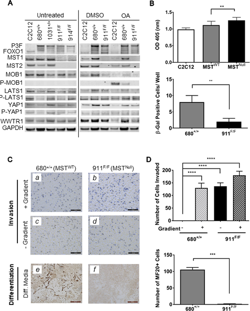
Figure 2. Tumor-derived cell lines provide insight into consequences of MST loss-of-function.

(A) Immunoblot of tumor-derived cell line lysates. Okadaic acid (OA, 150nM) was used as a pharmacologic activator of MST to reveal changes in downstream signaling. (B) Upper: BrdU incorporation is higher in MSTNull versus MSTWT tumor-derived cells. Data shown represents pooled values from three experiments including two cell lines each of MSTWT and MSTNull cell lines. Lower: MSTWT (680+/+) cells show increased β-Gal staining as compared to MSTNull (911F/F), indicating an increased number of senescent cells. Data shown represents four technical replicates. (C) Upper panels: representative images of the invasion potential of (a,c) MSTWT and (b,d) MSTNull cells. Note that MSTNull cells were able to invade even in the absence of a nutritional gradient, as shown by violet staining in (d). Lower panels: as evidenced by MF20 staining, (e) MSTWT cells were able to differentiate down the myogenic lineage, while (f) MSTNull cells were not. Data shown represents four technical replicates. (D) Quantitation of invasion (upper panel) and differentiation (lower panel) assays shown in (C).
