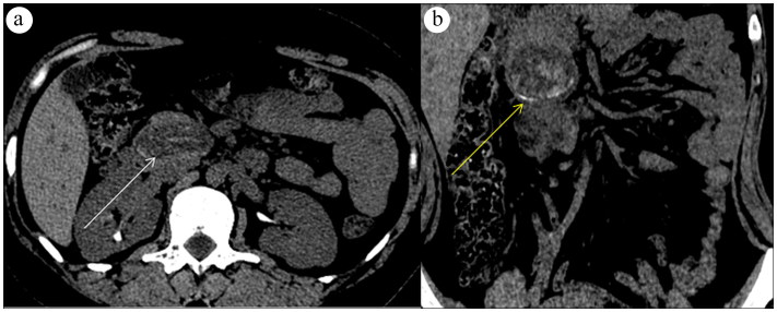
Figure 1.
(a) CT scan with intravenous contrast demonstrates a 4.0-cm hypodense mass in the pancreatic head and uncinate process (white arrow). (b) There is the suggestion of hyperdense/enhancing curvilinear densities representing either hyperdense/calcified septa or enhancement (arrow), because no precontrast imaging was performed.

