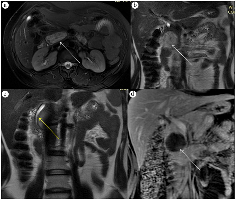
Figure 2.
MRI of the abdomen with and without intravenous contrast shows (a, b) the mass appearing heterogeneously T2 hyperintense (T2-weighted images, white arrows), causing (c) splaying and mild attenuation of the common bile duct (T2-weighted image, arrow). (d) Postcontrast image demonstrates no enhancement (white arrow).

