
Figure 1. Brain magnetic resonance imaging (MRI) of a patient with metachromatic leukodystrophy (MLD).
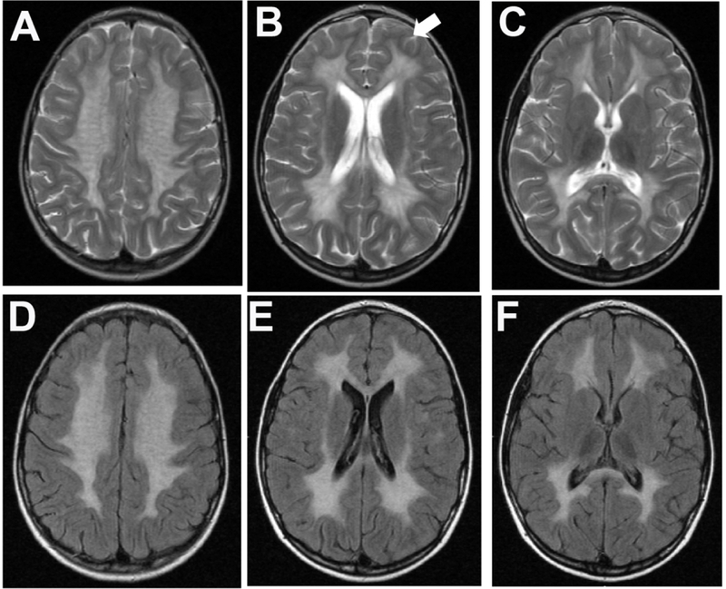
A 7-year old female who had one-year history of progressive ataxia, cognitive impairment and dysarthria which started approximately 18 months before the brain MRI was performed. She was diagnosed with the juvenile form of MLD. Sequences of fluid attenuated inversion recovery (FLAIR; A-C) and T2-weighted turbo spin echo (T2W_TSE; D-F) of transverse sections are shown. (A) The brain MRI shows typical widespread hyperintensity signals affecting the entire centrum semiovale of both hemispheres, frontal, parietal and occipital regions extending bilaterally to the temporal white matter. In panel B, the white-arrow shows subcortical U-fibers in the L frontal lobe, that are initially spared in MLD. (C and F) Brainstem shows small scattered areas of punctate high-signal representing also white matter demyelination, mostly in the thalami and subthalamic regions in the T2W and FLAIR sequences.